Skin. Signals. Sensation.
Follow the signal.
1. Detect a pattern
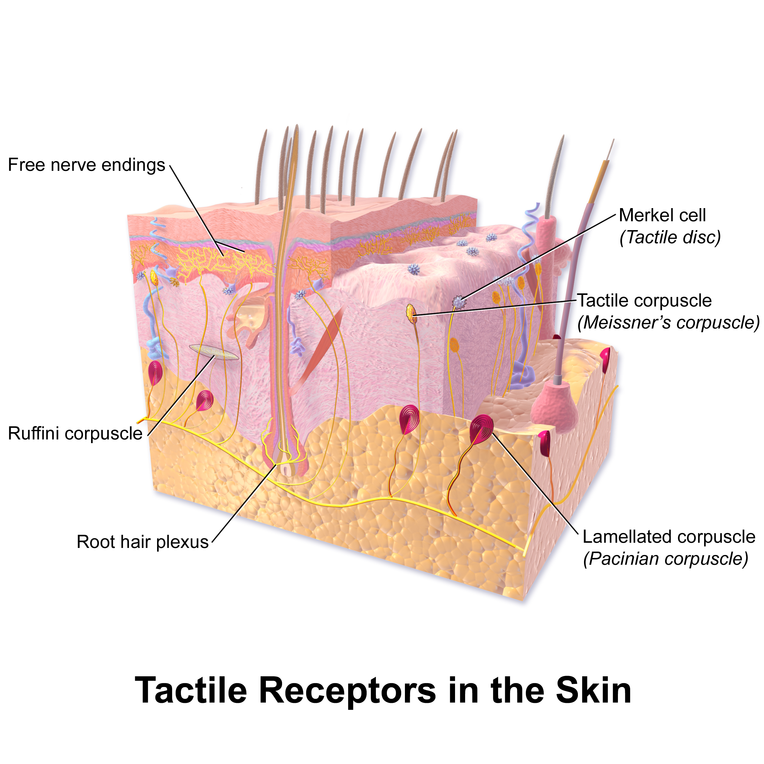 Open larger labeled diagram ↗
- Merkel / SA-I
- Held indentation and fine detail
- Meissner / RA-I
- Changing contact and flutter
- Pacinian / RA-II
- High-frequency vibration
- Ruffini / SA-II
- Skin stretch; traditional anatomical pairing
Four classic tactile afferent types in hairless skin. Slowly adapting responses can continue during a held stimulus. Rapidly adapting responses emphasize change.
2. Convert force into a signal
Skin deforms
Mechanically gated channels open
Net positive current changes local voltage
Threshold triggers action potentials
PIEZO2 is a major touch channel. A graded local voltage change can start all-or-none traveling spikes.
3. Follow the main routes
Right / left = body sides
Fine touch / vibration
Pain / temperature
- Fine touch / vibration
- Crosses in the medulla
- Pain / temperature
- Crosses in the spinal cord
These are two major pathways from the body. They relay toward the thalamus and sensory cortex.
4. Compare temperature and pain

- TRPM8
- Cold and menthol
- TRPV1
- Noxious heat and capsaicin
- Aδ nociceptors
- Thinly myelinated; typically faster, sharper
- C nociceptors
- Unmyelinated; typically slower, burning or aching
Warmth depends on activity across neurons. Nociception processes potentially harmful stimulation. Pain is the personal experience.
McKemy et al., 2002; Caterina et al., 1997; Paricio-Montesinos et al., 2020; Dubin & Patapoutian, 2010; IASP, 2020
5. Notice what can change
Transmission can change
- Gate control
- Touch input can recruit inhibitory spinal circuits.
- Endorphins
- Opioid receptor signaling can reduce transmitter release and excitability.
- Descending control
- Brain signals can inhibit or facilitate transmission.
- Phantom sensation
- A sensation can be located in an absent limb.
Phantom sensations may be painless. Phantom pain is felt in the absent limb; residual-limb pain is in remaining tissue. Peripheral, spinal, and brain processes can interact.
Mendell, 2014; Che & Roth, 2023; Heinricher et al., 2009; Hanyu-Deutmeyer et al., 2023
6. Try tap, hold, glide
- Tap your opposite palm gently
- Hold comfortable contact for 20 seconds
- Glide a fingertip a short distance
Notice contact starting, maintained pressure, and movement. Observe instead if you prefer. Use light contact on intact skin. This does not test individual receptors.
Which felt most obviously changing? Could you still sense the held contact?
Full APA references and image credits
Download APA references PDF ↗- Dougherty, P. (2020, October 7). Chapter 2: Somatosensory systems (C. Tsuchitani, content contributor). In Neuroscience online. McGovern Medical School at UTHealth Houston. https://nba.uth.tmc.edu/neuroscience/m/s2/chapter02.html Open source
- Dougherty, P. (2026, July 7). Chapter 4: Somatosensory pathways (C. Tsuchitani, content contributor). In Neuroscience online. McGovern Medical School at UTHealth Houston. https://nba.uth.tmc.edu/neuroscience/m/s2/chapter04.html Open source
- Betts, J. G., Young, K. A., Wise, J. A., Johnson, E., Poe, B., Kruse, D. H., Korol, O., Johnson, J. E., Womble, M., & DeSaix, P. (2022). Anatomy and physiology 2e. OpenStax. https://openstax.org/books/anatomy-and-physiology-2e/pages/1-introduction Open source
- Paré, M., Behets, C., & Cornu, O. (2003). Paucity of presumptive Ruffini corpuscles in the index finger pad of humans. The Journal of Comparative Neurology, 456(3), 260–266. https://doi.org/10.1002/cne.10519 Open source
- Chesler, A. T., Szczot, M., Bharucha-Goebel, D., Čeko, M., Donkervoort, S., Laubacher, C., Hayes, L. H., Alter, K., Zampieri, C., Stanley, C., Innes, A. M., Mah, J. K., Grosmann, C. M., Bradley, N., Nguyen, D., Foley, A. R., Le Pichon, C. E., & Bönnemann, C. G. (2016). The role of PIEZO2 in human mechanosensation. The New England Journal of Medicine, 375(14), 1355–1364. https://doi.org/10.1056/NEJMoa1602812 Open source
- McKemy, D. D., Neuhausser, W. M., & Julius, D. (2002). Identification of a cold receptor reveals a general role for TRP channels in thermosensation. Nature, 416(6876), 52–58. https://doi.org/10.1038/nature719 Open source
- Caterina, M. J., Schumacher, M. A., Tominaga, M., Rosen, T. A., Levine, J. D., & Julius, D. (1997). The capsaicin receptor: A heat-activated ion channel in the pain pathway. Nature, 389(6653), 816–824. https://doi.org/10.1038/39807 Open source
- Paricio-Montesinos, R., Schwaller, F., Udhayachandran, A., Rau, F., Walcher, J., Evangelista, R., Vriens, J., Voets, T., Poulet, J. F. A., & Lewin, G. R. (2020). The sensory coding of warm perception. Neuron, 106(5), 830–841.e3. https://doi.org/10.1016/j.neuron.2020.02.035 Open source
- Dubin, A. E., & Patapoutian, A. (2010). Nociceptors: The sensors of the pain pathway. Journal of Clinical Investigation, 120(11), 3760–3772. https://doi.org/10.1172/JCI42843 Open source
- International Association for the Study of Pain. (2020, July 16). IASP announces revised definition of pain. https://www.iasp-pain.org/publications/iasp-news/iasp-announces-revised-definition-of-pain/ Open source
- Melzack, R., & Wall, P. D. (1965). Pain mechanisms: A new theory. Science, 150(3699), 971–979. https://doi.org/10.1126/science.150.3699.971 Open source
- Mendell, L. M. (2014). Constructing and deconstructing the gate theory of pain. Pain, 155(2), 210–216. https://doi.org/10.1016/j.pain.2013.12.010 Open source
- Heinricher, M. M., Tavares, I., Leith, J. L., & Lumb, B. M. (2009). Descending control of nociception: Specificity, recruitment and plasticity. Brain Research Reviews, 60(1), 214–225. https://doi.org/10.1016/j.brainresrev.2008.12.009 Open source
- Che, T., & Roth, B. L. (2023). Molecular basis of opioid receptor signaling. Cell, 186(24), 5203–5219. https://doi.org/10.1016/j.cell.2023.10.029 Open source
- Makin, T. R., Scholz, J., Filippini, N., Henderson Slater, D., Tracey, I., & Johansen-Berg, H. (2013). Phantom pain is associated with preserved structure and function in the former hand area. Nature Communications, 4, Article 1570. https://doi.org/10.1038/ncomms2571 Open source
- Vaso, A., Adahan, H.-M., Gjika, A., Zahaj, S., Zhurda, T., Vyshka, G., & Devor, M. (2014). Peripheral nervous system origin of phantom limb pain. Pain, 155(7), 1384–1391. https://doi.org/10.1016/j.pain.2014.04.018 Open source
- Hanyu-Deutmeyer, A. A., Cascella, M., & Varacallo, M. A. (2023, August 4). Phantom limb pain. In StatPearls. StatPearls Publishing. https://www.ncbi.nlm.nih.gov/books/NBK448188/ Open source
Image credits
- Hand photograph: cottonbro studio / Pexels. Original, Pexels license. Cropped for presentation framing.
- Skin diagram: BruceBlaus / Blausen.com staff (2014), Blausen 0809 Skin TactileReceptors. CC BY 3.0. Shown uncropped on this website. Cropped in the presentation to remove excess top whitespace and the bottom title band; all anatomical labels and leader lines preserved.
- Mint photograph: Ruby Sengar / Pexels. Original, Pexels license. Displayed with responsive framing on this website; shown uncropped in the presentation.
- Conceptual scientific diagrams: original teaching schematics, based on the cited sources. Response sketches are not measured recordings.